BACKGROUND
It is not easy for students to understand what is displayed in MR images, and how that depends on the sequence that was used. A teacher can explain it in a lecture or in a book, but students learn to understand complex relations best when they can change the input themselves, and observe how that affects the result. With this in mind, the program MRISIM has been constructed.
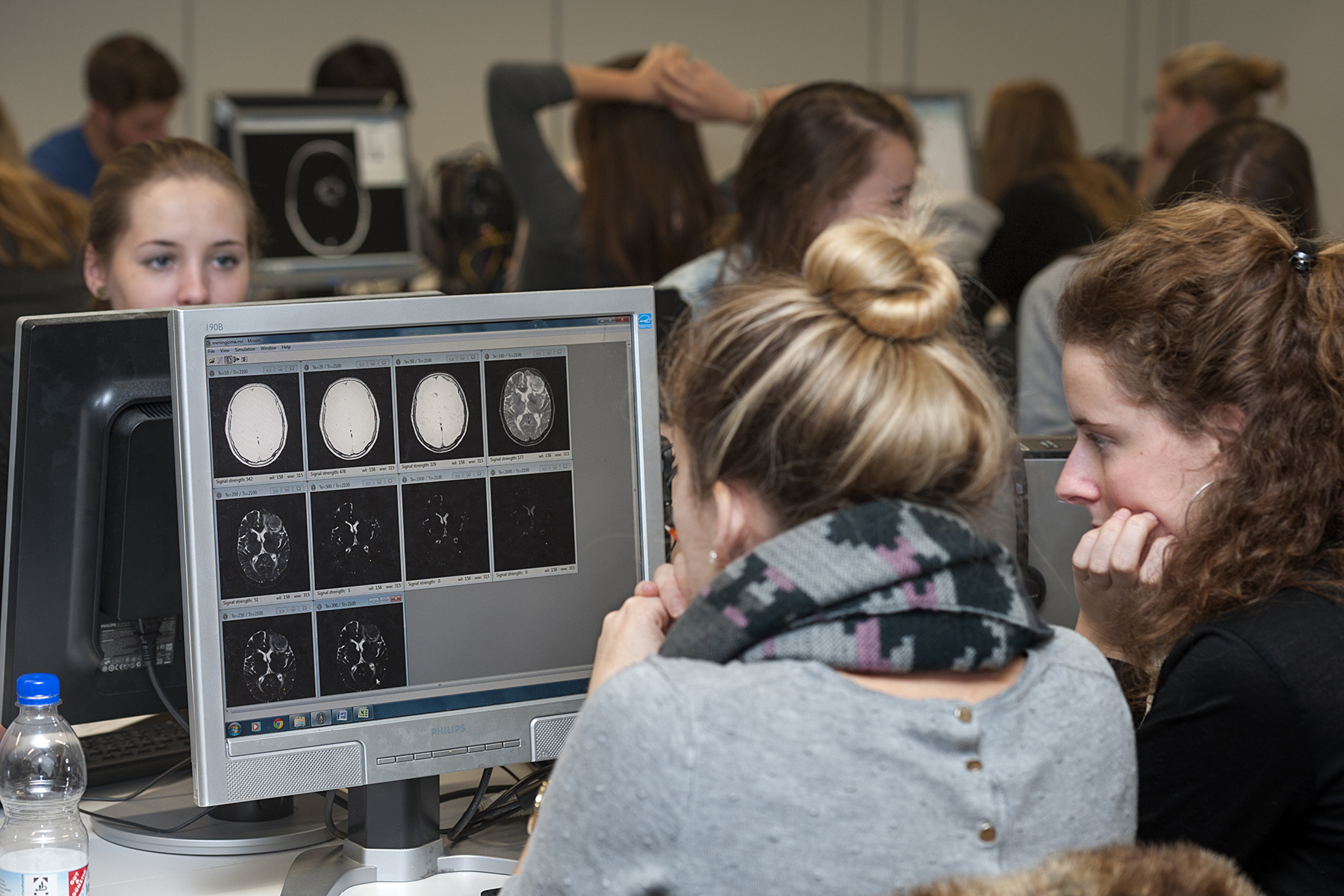
MRISIM provides the students with a virtual MR scanner, that produces MR images of real patients. The students can set the scanner parameters, such as TE and TR, and the program will compute and display the MR image for those settings.
The program can do this because its data files contain the T1, T2 and proton density value for each voxel (and in some data files additional properties such as DTI data). These properties were obtained by fitting them from a set of MR images of a single patient, recorded with different parameters.
At the Radboud University Medical Center, we have been using MRISIM for over a decade now, and found that it helps students to understand the essence of MR imaging. After an introductory lecture of 45 minutes and a computer class of 2 hours, students are able to answer exam questions where they have to choose MR settings for a specific purpose, or have to determine which settings were used in a given image.
There are also advanced classes, where MRISIM is used to teach students about more complicated sequenced such as FLAIR, or about diffusion weighted imaging and functional MRI.
We are interested in feedback from users. Also, we would like to extend our data collection. If you are interested in a colaboration, please contact us.